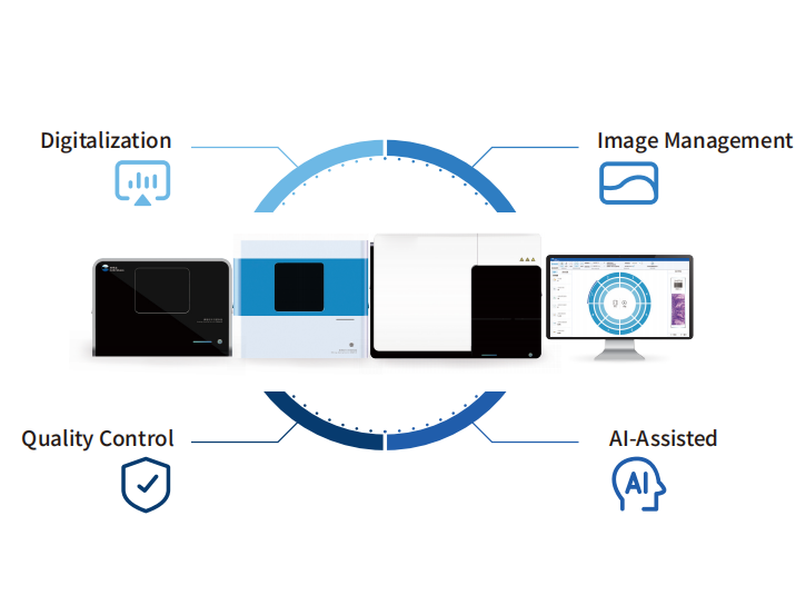
Hardware Agility: Bridging Diverse Scanning Ecosystems
The AI-powered diagnostic platform achieves robust interoperability with a vast spectrum of digital slide scanners, covering:
Global industry leaders: Philips, Zeiss, Leica Biosystems, Roche (ScanIm platform).
Niche specialists: 3DHISTECH (Europe), d-matrix (precision imaging), KFBIO (innovative optics).
Regional innovators: Motic, Unic
(Asia-Pacific), and more.
Crucially, it extends beyond third-party devices: we also support our own AI-optimized reagents, purpose-built scanners, and specialized microscopes. This means hospitals can either leverage existing infrastructure (preserving capital investments) or adopt tailored tools for enhanced performance—no forced hardware overhauls required.
System Integration: Syncing with Pathology IT Landscapes
On the software front, our AI-powered diagnostic platform integrates flawlessly with pathology information management systems (PIMS) and hospital IT ecosystems.
This eliminates manual bottlenecks:
Scanned slides flow directly into AI-driven analysis.
Pathologists’ diagnoses sync instantly with the PIMS.
Finalized reports auto-distribute to clinicians, embedded in the hospital’s core workflow.
Every step—from slide scanning to report delivery—operates as a unified, frictionless chain, cutting administrative delays and ensuring diagnostic insights reach care teams faster.

The Unified Advantage: Efficiency Without Compromise
By connecting hardware diversity, AI analysis, and system connectivity, we:
Streamlines workflows: Reduces manual handoffs, accelerating time-to-diagnosis.
Protects investments: Lets hospitals keep trusted devices while adding AI capabilities.
Empowers experts: Frees pathologists from repetitive tasks to focus on complex cases.
In short, we don’t just “add AI” to pathology—it weaves AI into the entire diagnostic journey, harmonizing with existing tools and systems to deliver faster, more consistent care. The future of pathology isn’t about replacing hardware or workflows—it’s about unifying them.
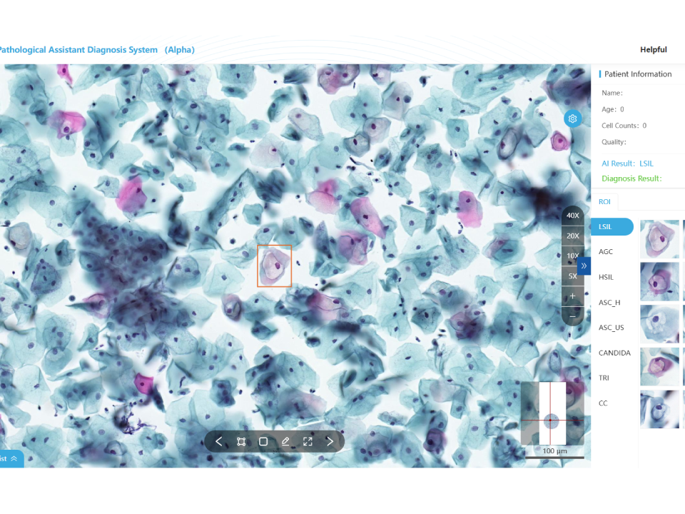
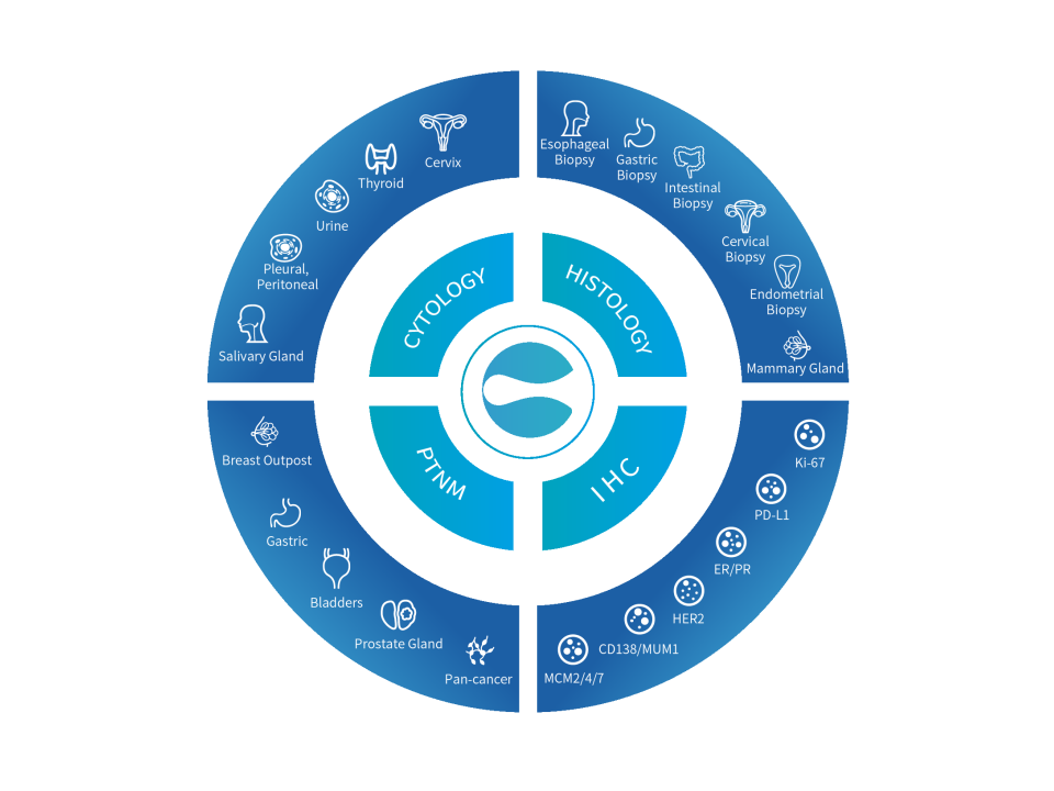
Cervical Cytology Assistant Diagnostic Module
Automatic recognition of 15 cell types. Accurate identification to reduce misdiagnosis. One-click report generation, dual-screen slide reading, interfacing with multiple systems. Validated by multi-center clinical trials: sensitivity ≥ 95%, specificity ≥ 85%. Whole slide analysis with TBS standard report. Customizable report template.
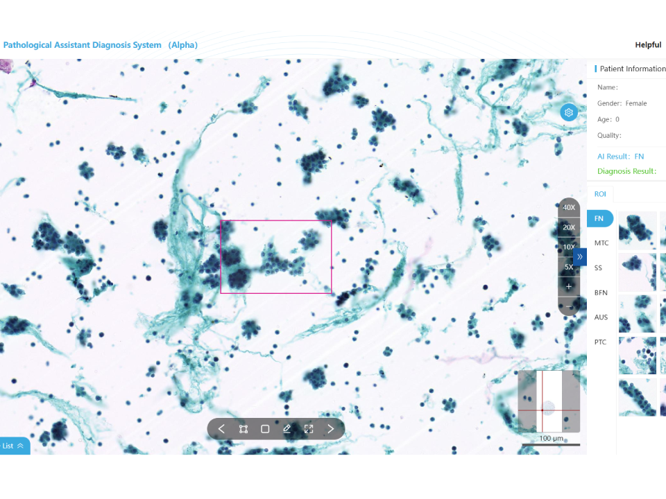
Thyroid Cytology Assistant Diagnostic Module
Automatic recognition of 6 cell types. High-speed analysis for improved efficiency. Validated by multi-center clinical trials: sensitivity ≥ 95%, specificity ≥ 90%. Whole slide analysis to reduce misdiagnosis TBS standardized pathology reports.
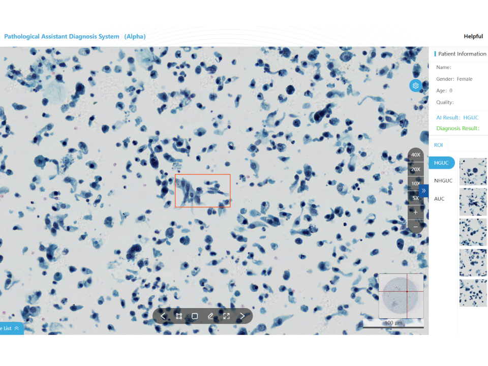
Urine Cytology Assistant Diagnostic Module
Automatic recognition of high-grade uroepithelial cancer cells Diagnostic report based on "The Evolution of the Paris System(TPS)" Validated by multi-center clinical trials: Sensitivity≥95%,Specificity≥93% Whole slide analysis to reduce missed defections Increase the positive detection rate
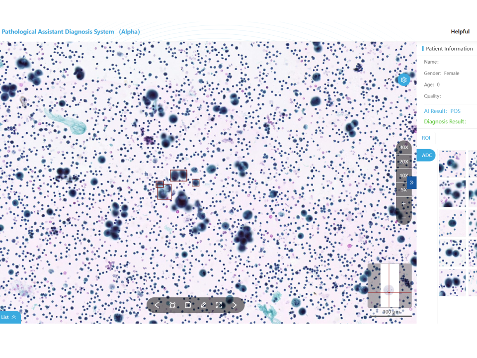
Hydrothorax and Ascites Cytology Assistant Diagnostic Module
Automatic recognition of malignant tumor cell Assist physicians with cytologic diagnosis. Whole slide analysis to reduce misdiagnosis.. Validated by multi-center clinical trials: sensitivity≥96%,specificity≥90%, Quantities 10000+ Increase the positive detection rate
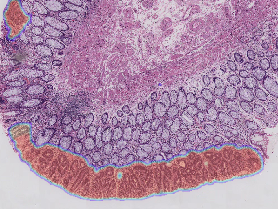
Gastric Biopsy Assistant Diagnostic Module
Efficient identification of tissue structural changes and cellular abnormalities. Automatically recognize eight lesion types,covering more than 95% of lesion types. Automatically capture the lesion area,effectively assist doctors in diagnosis. Combine pathology images, gastroscopy and medical history to improve physicians' diagnostic accuracy.
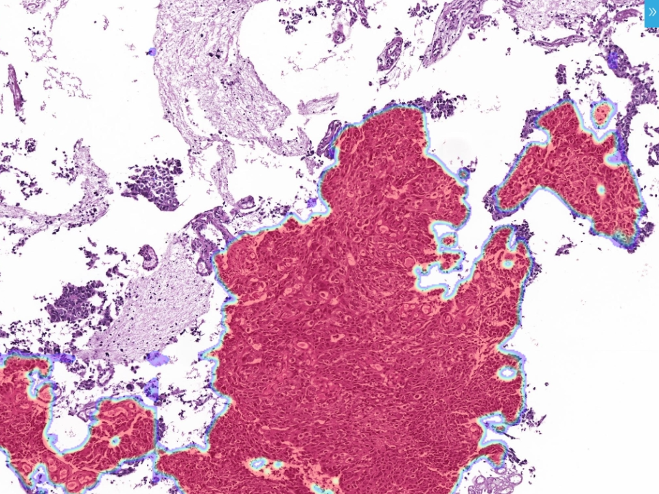
Intestinal Biopsy Assistant Diagnostic Module
Detection of small early lesion areas with high accuracy. Automatically recognize six lesion types,covering more than 95%of lesion types. Significantly shorten the diagnostic time and reduce workload. Combine pathology images,colonoscopy and disease history to improve diagnostic accuracy.

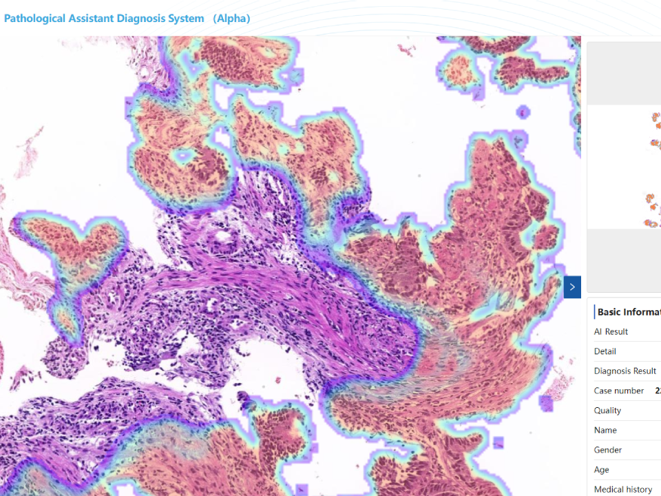
Cervical Biopsy Assistant Diagnostic Module
Whole slide analysis, automatic identification of squamous and adenocarcinomas Combine pathologicimages, colposcopy and medical history to improve diagnostic accuracy Combine diagnosis with cervical cytology diagnostic module,greatly improve the diagnostic accuracy of doctors.
Endometrial Biopsy Assistant Analysis Module
Fusion of high and low magnification models to discriminate tissue structural changes and cellular abnormalities. Rapid localization of lesions. Automatic recognition of six lesion types. Combining pathologic images and patient age to improve diagnostic accuracy.
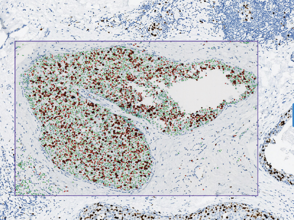
Immunohistochemistry Assistant Analysis Module
Whole slide analysis,intelligent cell counting. Algorithm intelligently segments the effective area to fit different diagnostic scenarios. Multiple staining images displayed on one screen for comprehensive comparative analysis. High concordance with the pathologist's interpretation with an accuracy of >0.94.
Cervical Cytology Assistant Diagnostic Module
Automatic recognition of 15 cell types. Accurate identification to reduce misdiagnosis. One-click report generation, dual-screen slide reading, interfacing with multiple systems. Validated by multi-center clinical trials: sensitivity ≥ 95%, specificity ≥ 85%. Whole slide analysis with TBS standard report. Customizable report template.