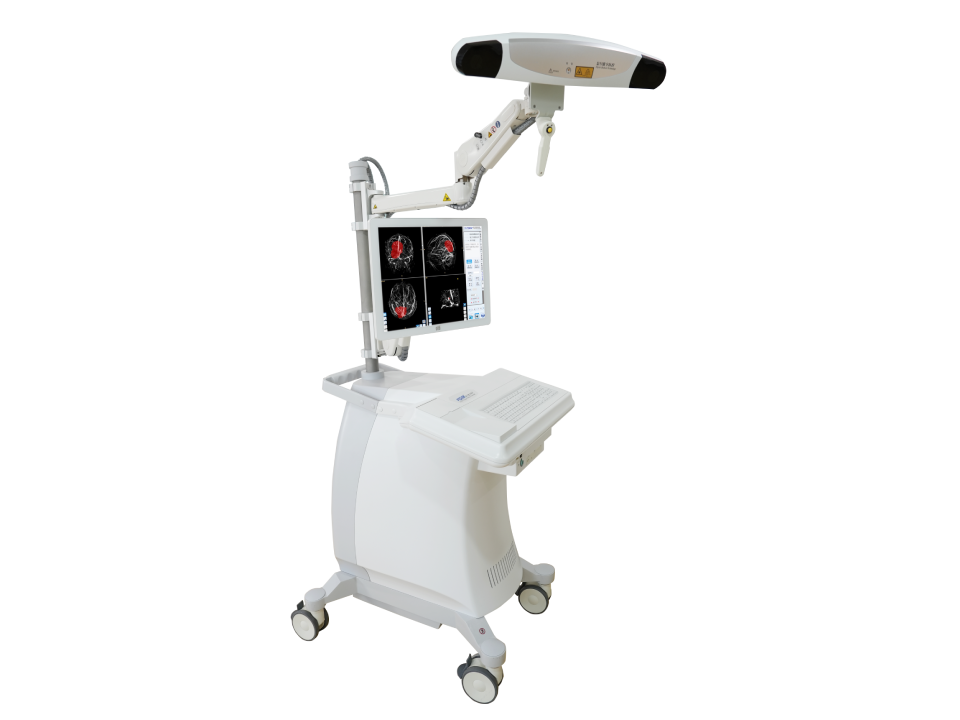
Excelim-04 Neurosurgical Stereotactic System

As a pioneer in digital medicine and computer-assisted surgery—the excelim-04 Neurosurgical Stereotactic System is an advanced integrated platform designed to elevate neurosurgical precision, minimize invasiveness, and enhance clinical outcomes. Merging cutting-edge optical positioning technology, AI-driven image processing, and multimodal data integration, this system empowers surgeons with real-time, 3D-guided navigation across a spectrum of complex neurosurgical procedures.

Excelim-04 Neurosurgical Stereotactic System

Core Fetures

All-in-One Design with High-Accuracy Optical Positioning

Multimodal Image Fusion & Intelligent Processing

At its core lies a high-precision optical tracking system that delivers sub-millimeter positional accuracy, ensuring real-time alignment of surgical instruments with pre-operative imaging. This seamless integration eliminates delays, keeping surgeons focused on the procedure while maintaining pinpoint accuracy—critical for delicate operations near functional brain regions or vital vasculature.

Leveraging Decision Level Intelligent Image Fusion Technology, the excelim-04 integrates diverse imaging modalities (CT, MRI, DTI, MRA, fMRI, CTA, DSA) into a unified 3D visualization. This fusion clarifies anatomical relationships, enhances lesion detectability, and provides a comprehensive view of critical structures like fiber bundles (e.g., pyramidal tracts) via automated MR-DTI processing. Surgeons gain unprecedented clarity to avoid damage to functional areas.

Versatile Registration & Navigation Modes

Advanced 3D Reconstruction & Lesion Analysis

The system supports multiple registration methods to adapt to varied clinical scenarios, paired with diversified navigation modes tailored to specific procedures—from biopsy to tumor resection. This flexibility ensures optimal performance whether for routine cases or complex interventions requiring customized workflows.

Transforming 2D medical images into vivid 3D models, the excelim-04 enables surgeons to visualize lesions and surrounding anatomy in intricate detail. Features like automated lesion segmentation, volume calculation, and MIP-based vascular imaging further streamline pre-operative planning, ensuring precise surgical strategies.

Comprehensive Toolkit for Minimally Invasive Procedures

Equipped with specialized accessories—including sterile probes, reference frames, surgical instrument adapters, and puncture components—the system supports minimally invasive techniques such as frameless biopsy, cerebral hemorrhage drainage, and ventricular endoscopy. A dedicated sterilized equipment box ensures compliance with intraoperative safety standards.

Clinical Applications

Frameless Puncture Biopsy

Enables precise, minimally invasive tissue sampling with instrument adapters that align with navigation guidance, reducing procedural risk.

Brain Tumor Resection

Real-time monitoring of lesion boundaries and adjacent critical structures (e.g., blood vessels, fiber tracts) minimizes collateral damage, preserving neurological function.

Cerebral Hemorrhage Drainage

Automatically outlines hemorrhage zones, calculates volumes, and suggests optimal trajectories, with real-time navigation to ensure accurate catheter placement.

Endoscope & Ventriculoscope Integration

Fuses dynamic endoscopic footage with static navigation images, providing surgeons with contextualized, real-time visual feedback during intraventricular procedures.

Functional Neurosurgery

Utilizes MR-DTI fiber tract mapping to preserve motor, sensory, or language pathways, critical for operations in eloquent brain regions.
Ready to transform your neurosurgical practice? Contact us to learn how excelim-04 can elevate your team’s capabilities—from pre-operative planning to intra-operative execution.

Interested in Talking with a Medical Expert?

Contact us today to learn more.