3D Intelligent Interactive Surgical Planning System

The Gmedisys 3D Intelligent Interactive Surgical Planning System is a digital medical solution based on holographic 3D medical imaging technology. Its core lies in 3D reconstruction from 2D images, intelligent interaction, and visual navigation, providing full-process support for surgeries across multiple departments. Its basic functions include:

  • Converting multimodal images such as CT, MRI, and endoscopy into precise 3D models, clearly presenting the anatomical relationships between lesions, blood vessels, and surrounding tissues.
  • Supporting preoperative planning (e.g., determining resection ranges, simulating surgical paths), intraoperative navigation (real-time comparison between models and operations), doctor-patient communication (visual explanation of conditions), and scientific research & teaching (building 3D databases).
  • Cooperating with the AQI Digital Intelligent Interaction System, realizing spatial interaction with 3D models through VR devices, 3D glasses, and styluses, enhancing 3D immersion, and supporting multi-person collaboration and multi-angle observation.

Applications in Department-Specific Surgeries

Hepatobiliary Surgery

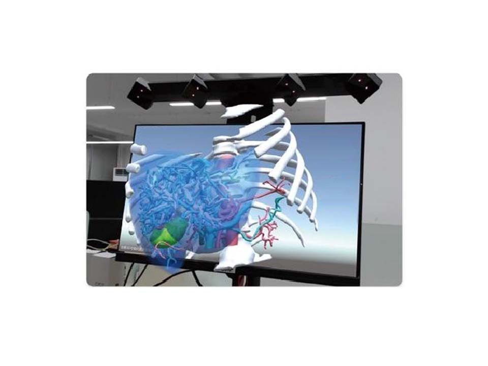
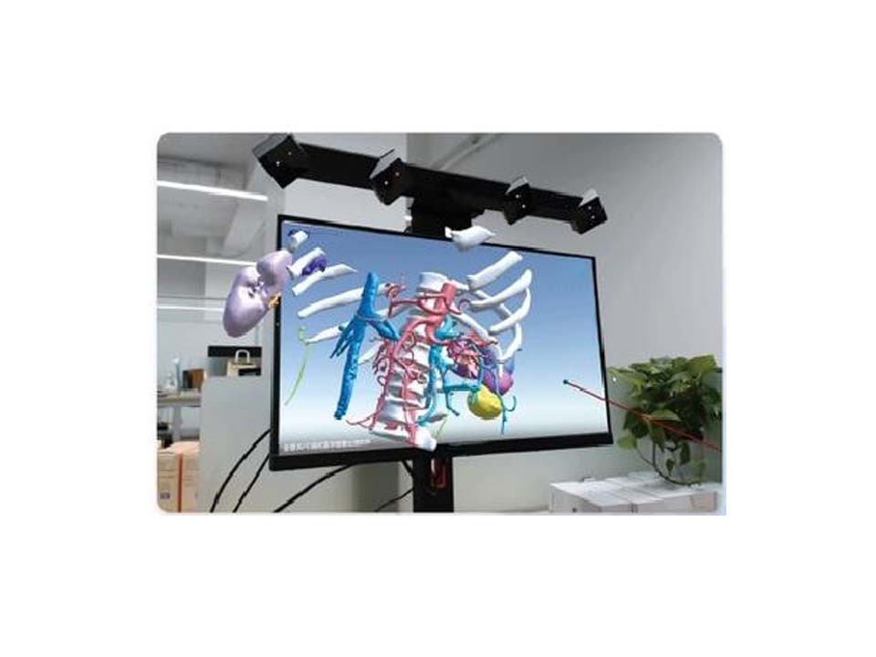
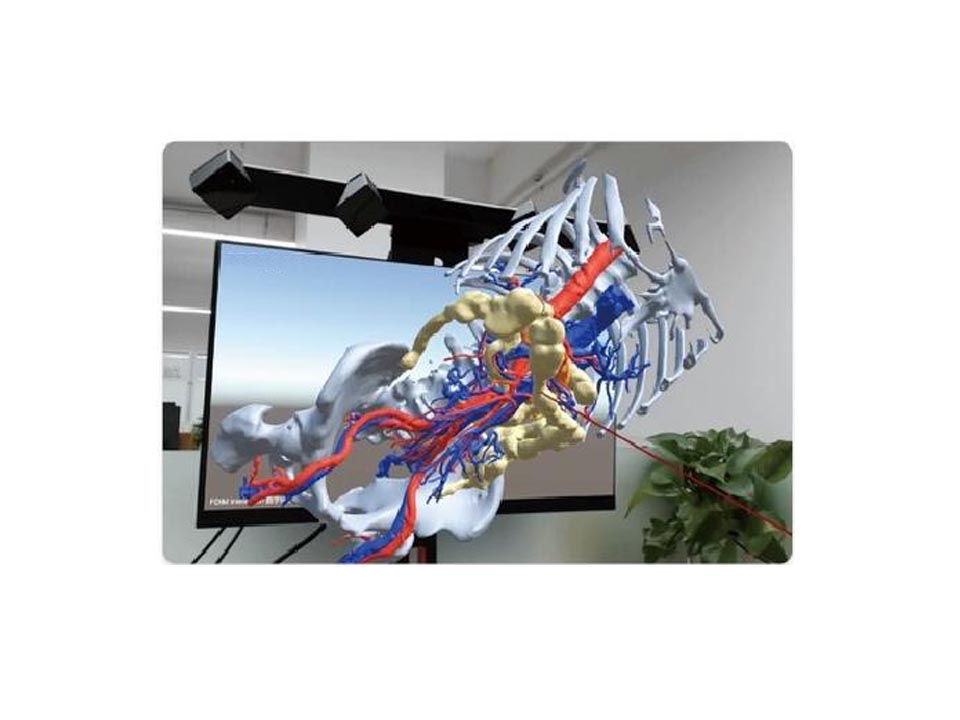
Performing liver segment segmentation based on liver watershed analysis, expanding the lesion by 1cm to determine the safe margin, clarifying the positional relationship between tumors and portal veins, hepatic veins, and hepatic arteries, and planning the watershed resection range.


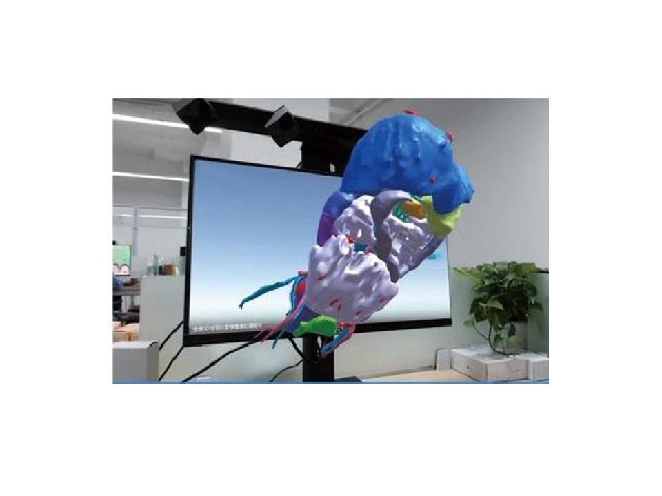
Precisely marking liver segments, focusing on displaying the association between blood vessels and lesions, and supporting liver segment navigation and data measurement.


Improving the accuracy of liver resection surgeries, reducing the risk of vascular injury and bleeding, lowering the probability of tumor residue, and shortening the time for preoperative plan formulation.

Gynecological Surgery

Reconstructing organs and lesions through pelvic MRI, evaluating tumor ranges and blood supply sources (e.g., uterine arteries), simulating surgical paths, and measuring the distance between tumors and important organs.


Clearly identifying complex pelvic anatomical structures, supporting personalized plan customization, and optimizing paths according to surgeons’ operating habits.


Reducing damage to normal tissues, preserving reproductive function, lowering the risk of complications, and improving the efficiency of doctor-patient communication through visual models.

Urinary Surgery

Adopting the “2mm spacing method” to plan nephron-sparing surgeries for kidney tumors, analyzing the relationship between tumors and renal arteries/veins, and simulating resection ranges.


Supporting fine cutting simulation and real-time 2D/3D comparison, and displaying models on multiple platforms to assist intraoperative navigation.


Maximizing the preservation of healthy renal tissue, reducing the risk of renal function damage, shortening surgical time, and improving surgical safety.

Gastrointestinal Surgery

Converting 2D images into 3D models of gastrointestinal lesions, displaying lesion morphology and blood supply, planning surgical paths, and comparing preoperative and postoperative efficacy.


Integrating multimodal images to generate standardized reports, supporting dynamic adjustment of intraoperative models and saving of key interfaces.


Improving the accuracy of tumor resection, reducing complications such as anastomotic leakage, optimizing surgical processes, and providing an intuitive basis for efficacy evaluation.

Thoracic Surgery

Reconstructing 3D models of pulmonary nodules (including 5th-level vascular branches), planning wedge or subsegment resection ranges, and simulating the disconnection paths of blood vessels and bronchi.

Supporting subsegment-level segmentation, non-invasive virtual positioning, and adapting to early lung cancer and complex multi-nodule cases.

Preserving more lung function, shortening the time for intraoperative lesion localization, reducing surgical trauma, and improving the success rate of high-difficulty surgeries.

Advantages

Technical Advantages

Personalized 3D reconstruction, integration of multimodal information, AI-assisted automated measurement, and cross-platform interaction (supported by the AQI system), adapting to surgical needs of various departments.

Clinical Values

Improving surgical accuracy and safety, reducing tissue damage and complications; alleviating patients’ psychological pressure through visual communication, and accelerating postoperative recovery.

Scientific Research and Teaching

Supporting the construction of 3D organ databases, facilitating multi-center research and clinical transformation, and providing stereoscopic anatomical teaching tools for medical students.

Support from 3D Digital Medicine Center

The system relies on the 3D Digital Medicine Center, integrating holographic surgical planning, navigation, and 3D printing technologies, to provide a full-process “preoperative-intraoperative-postoperative” solution for various departments. It promotes the application of digital technologies in clinical diagnosis, treatment, and scientific research, realizing the precision, personalization, and remoteness of medical services.

Ready to elevate your surgical precision and patient care? Reach out today to explore how Gmedisys’s intelligent solutions can transform your clinical practice across hepatobiliary, gynecological, urinary, gastrointestinal, and thoracic surgeries.

Interested in Talking with a Medical Expert?

Contact us today to learn more.